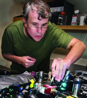
Earlier this month Eric Betzig shared the Nobel Prize in chemistry for his work on high-resolution microscopes -- specifically the one he'd designed and built on a friend's living room floor.
But when Betzig, a researcher at the Howard Hughes Medical Institute's Janelia Research Campus in Ashburn, Virginia, got news of his win, his best work yet was still a few weeks away from being published. Thursday in Science, he and a team of his colleagues reported on a new microscopy technique that allows them to observe living cellular processes at groundbreaking resolution and speed.
But when Betzig, a researcher at the Howard Hughes Medical Institute's Janelia Research Campus in Ashburn, Virginia, got news of his win, his best work yet was still a few weeks away from being published. Thursday in Science, he and a team of his colleagues reported on a new microscopy technique that allows them to observe living cellular processes at groundbreaking resolution and speed.
Betzig came up with his Nobel-winning microscope (PALM) when he'd grown frustrated with the limitations of other microscope technologies. The so-called lattice light-sheet microscopy that he describes in Thursday's paper was the result of his eventual boredom with PALM.
"Again, I just started to understand the limits of the technology," Betzig said. PALM was great at looking at living systems, but only when they moved slowly. It couldn't take measurements quickly enough to get high-resolution pictures of fast cellular divisions.
 Trying to understand biology using these microscopes is like piecing together a football game from high-resolution photos, Betzig said: You can see images of a pass, and a touchdown, and of the cheerleaders doing a pyramid. But the rules of the game would only become clear once you saw a game on video.
Trying to understand biology using these microscopes is like piecing together a football game from high-resolution photos, Betzig said: You can see images of a pass, and a touchdown, and of the cheerleaders doing a pyramid. But the rules of the game would only become clear once you saw a game on video.
"I'd been looking at those pictures my whole life," Betzig said. "It was time to take a look at the living stuff in action."
Until now, the best microscope for viewing living systems as they moved were confocal microscopes. They beam light down onto a sample of cells. The light penetrates the whole sample and bounces back.
But even though a scientist can only focus his lens on one small section of the sample, light is being blasted onto the cells from above and below. This causes two problems: It creates a sort of haze around the area being focused on, and it's also damaging to the cell sample. The light is toxic, and degrades the living system over time.
Betzig's new microscope solves this by generating a sheet of light that comes in from the side of the sample, made up of a series of beams that harm the sample less than one solid cone of light. Scientists can now snap a high-res image of the entire section they're illuminating, without exposing the rest of the sample to any light at all.
The researchers involved have presented videos of 20 different biological processes in unprecedented clarity, and that's just a small sampling of what the technique can accomplish. By observing the way cells truly behave as they divide and grow, Betzig said, we'll have a better shot at understanding the causes and development of cancer, as well as how different congenital problems develop as embryos experience cell division.
Harvard professor of cell biology Tomas Kirchhausen wasn't involved in the new microscope's development, but he was blown away when he saw it in action at Betzig's lab last year.
"I was so impressed by the instrument and its potential capabilities that I asked if it would be possible to clone it for my own lab," Kirchhausen said. "And Eric generously agreed."
Kirchhausen's version of the microscope has been functional since August. For the work his lab does looking at living cells and tissues, it's been "transformative," he said.
For the first time, his group was able to get accurate measurements as they observed cells changing shape and size while undergoing division. "This simple question of whether cells change their volume and surface area when they change shape to divide just wasn't possible to study before. This works fantastically," he said.
The microscope allows his group to see cellular biology unfold in the context of real cells and tissues, Kirchhausen said -- instead of on glass slides.
Betzig is eager to get his new microscope into as many hands as possible, and believes that this development will have more of an impact on biological research than the work that earned him a Nobel Prize.
"Every week we have new research groups coming in," he said, "and not to pat my own back too much, but I feel a bit like Galileo -- everywhere you point this thing, you're going to learn something new."
"Every week we have new research groups coming in," he said, "and not to pat my own back too much, but I feel a bit like Galileo -- everywhere you point this thing, you're going to learn something new."
But Betzig already has hopes of moving past the groundbreaking new tech. Lattice light sheet microscopy is advanced, but it still has its limits: Like all light-based microscopes, it can only take clear images of the surface of an object.
"The eventual goal is to marry all of my work together to make a high-speed, high-resolution, low-impact tool that can look deep inside biological systems," Betzig said.
And he certainly has the faith of his scientific peers. "It's great that we could clone the microscope for my lab," Kirchhausen said, "But I wish we could clone Betzig, too."
No comments:
Post a Comment